Esophageal-Tracheal- Combitube ® (ETC)
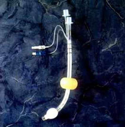
The Combitube is a twin lumen device designed for use in emergency situations and difficult airways. It can be inserted blindly into the oropharynx and usually enters the esophagus. It has a low volume inflatable distal cuff and a much larger proximal cuff designed to occlude the oro- and nasopharynx..
If the tube has entered the trachea, ventilation is achieved through the distal lumen as with a standard ETT. More commonly the device enters the esophagus and ventilation is achieved through multiple proximal apertures situated above the distal cuff . In the latter case the proximal and distal cuffs have to be inflated to prevent air from escaping through the esophagus or back out of the oro- and nasopharynx..
The Combitube can only be used in the adult population as no pediatric sizes are available. Complications of the Combitube include an increased incidence of sore throat, dysphagia and upper airway hematoma when compared to endotracheal intubation and LMA.. Esophageal rupture is a rare complication but has been described .Known esophageal disease is a contra-indication to the use of the Combitube. These complications may be partially preventable by avoiding over-inflation of the distal and proximal cuffs (see recommendations below). Compared to intubation with an endotracheal tube under direct laryngoscopy or using the LMA, the Combitube seems to exert a more pronounced hemodynamic stress response.
Although it is possible to maintain an airway with the Combitube, endotracheal intubation is the preferred method for definitively securing the airway. Either the oral or the nasal route can be used for fiberoptic-guided airway exchange. The Combitube is left in place and the proximal cuff is partially deflated for fiber-optic intubation with an endotracheal tube.
Little preparation is needed beyond testing both cuffs for leaks . The pilot balloon of the distal cuff is white and is marked with the number 2. Test the distal cuff by inflating with 15 ml of air. The pilot balloon of the proximal cuff is blue and is marked with the number 1. Test the proximal cuff by inflating with 85 ml of air..
The available sizes are 41 Fr and 37 Fr. The original recommendation by the manufacturer is to use 41 Fr for patients taller than 152 cm and 37 Fr for patients below that height. However, the bulky design of the 41 Fr can make it more technically difficult to insert and some authors have reported satisfactory results using the 37 Fr Combitube on taller patients. A redesigned Combitube has been described by creating an enlarged hole in the pharyngeal lumen that allows fiberoptic access, tracheal suctioning, and tube exchange over a guide wire.
The Combitube® can be inserted blindly without the aid of a laryngoscope . However, use of a laryngoscope has been reported to facilitate placement of the Combitube, it appears that the laryngoscope aids insertion by forcefully creating a greater space in the hypopharynx.
- Induce patient as if for regular intubation.
- Patient head position can be neutral.
- When direct laryngoscopy is attempted and the vocal cords can be visualized, the Combitube should be placed in the trachea and used as a regular endotracheal tube.
- Inflate the distal cuff with just enough air until no leak is present.
- Check for bilateral breath sounds over the lungs and confirm endotracheal placement on the capnogram.
- Connect the breathing circuit to the white connector number 2.
- If the Combitube is placed blindly, the left hand should elevate the chin while the right hand maneuvers the Combitube. Alternatively, more space can be created in the hypopharynx by using a laryngoscope with the left hand. The Combitube should be inserted to such a depth that the upper incisors are between the two black guidelines on the external surface of the tube:
- Inflate the distal cuff with 12 ml.
- Ventilate through the white connector number 2 and listen for gurgling sounds over the epigastrium or breath sounds over the lungs. If breath sounds are heard over the lungs the Combitube has been placed in the trachea and can be used as a regular ETT as described above after confirmation on the capnogram. If gurgling sounds are heard over the epigastrium, the Combitube is located in the esophagus.
- Inflate the proximal cuff with just enough air until either no leak is present or a subjective sensation of increased resistance to cuff inflation is encountered. This is usually achieved by inflating with 50-75 ml of air. This is less than the 85 ml recommended by the manufacturer but has been found to cause less upper airway trauma (1)
- Ventilate through the blue connector number 1, listen for breath sounds over the lungs and confirm ventilation on the capnogram..
Unable to ventilate patient through blue connector number 1
Make sure the Combitube is not per chance in the trachea. Attempt to ventilate through connector number 2, if breath sounds are heard over the lungs then the combitube has been placed in the trachea instead of the esophagus. Deflate the large proximal pharyngeal cuff and use the Combitube as a regular ETT.
Unable to ventilate patient through either connector
Confirm that the combitube has been placed in the esophagus by listening for epigastric gurgling sounds while ventilating through connector number 2. Then withdraw the combitube 2-3 cm at a time while ventilating through connector number 1 until breath sounds are heard over the lungs. The most common cause of this inability to ventilate to ventilate through either connector is an excessive insertion depth of the combitube (relative to the patient). This will cause obstruction of the glottic opening by the large proximal pharyngeal cuff (1,5).
Dr Michael Frass, Inventor, presents a video in English at http://www.combitube.org/
There's an introduction video by : Dr. Alan D. Kaye, Chairman and Professor of Anesthesia, Texas Tech University, USA and a Live Combitube Intubation Video as well by Dr. Peter Krafft, Dept of Anesthesiology, University of Vienna .
An Spanish web may be found at http://www.akh-wien.ac.at/combitube/spanisch.html .
Another superb explanation made by the Kendall-Sheridan corporation may be found at http://www.anest.ufl.edu/~eduweb/airway/combitube/combitube.html .
The AC-537 Combitube®, 37 French costs U$S 51.
Video at: http://www.laerdal.no/simman/videoIntubation.htm |